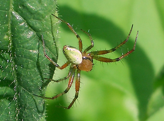
Male specimen. Specimen photographed in
Göttingen (Niedersachsen, Germany) on
June 01, 2008.

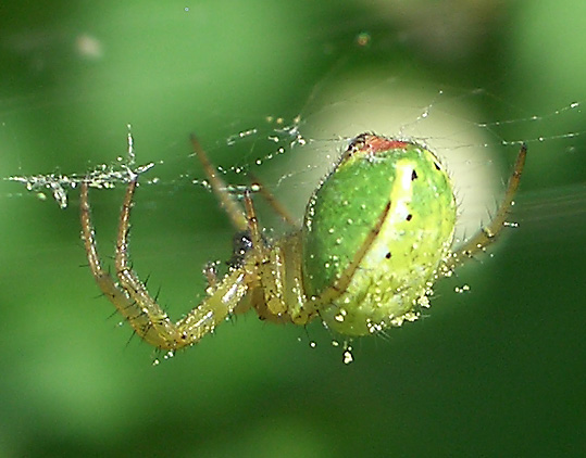
Female specimen. Specimen photographed in
Göttingen (Niedersachsen, Germany) on
June 01, 2008.

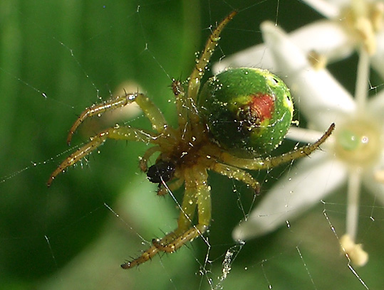
Female specimen.
Specimen photographed in Göttingen
(Niedersachsen, Germany) on June 01, 2008.

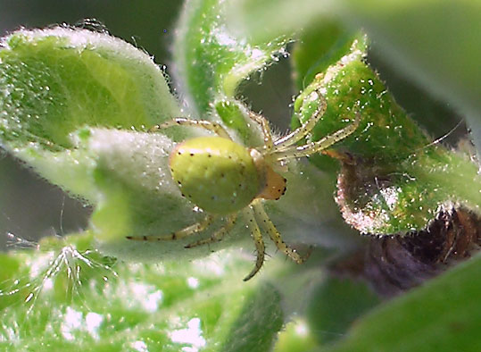
Female specimen.
Specimen photographed in Göttingen
(Niedersachsen, Germany) on May 11, 2008.
Subspecies
Original description
Synonyms
Araneus
cucurbitinus Clerck, 1757
Araneus cucurbitinus cucurbitinus Clerck,
1757
Araniella
cucurbitina (Clerck, 1757)
Araniella
cucurbitina cucurbitina (Clerck, 1757)
Aranea cucurbitina (Clerck,
1757)
Aranea
cucurbitina
cucurbitina (Clerck, 1757)
Epeira cucurbitina (Clerck,
1757)
Miranda cucurbitina (Clerck,
1757)
Aranea frischii
Scopoli, 1763
Aranea octopunctata Linnaeus, 1767
Aranea viridispunctata De Geer, 1778
Aranea depressa Razoumowsky, 1789
Epeira squamosa Seidel, 1849
Epeira cossoni Simon, 1885
Araneus cossoni
(Simon, 1885)
Araneus
cucurbitinus typicus Kulczynski, 1905
Identification
Araniella
cucurbitina and Araniella opisthographa are
extremely similar. To my knowledge, the females
cannot be separated with confidence. There is a
slight difference in the length of the scapus of
the female genitalia, but this is difficult to see
on a single individual and is only becoming
obvious if larger series are available. The males
differ in their palp morphology. Araniella
cucurbitina has a short median apophysis with a
kink that gives the apophysis a hook-shaped
appearance, whereas this apophysis is long, and
smoothly curved along its entire length in
Araniella opisthographa. In addition, the hair
pattern on the ventral side of the femur of the
first walking leg differs between the two species.
The males of Araniella cucurbitina have a clear
gap in the hair pattern, usually only 3 basal and
1 apical hair, whereas Araniella opisthographa has
a pattern of 7 (sometimes 8) hairs, regularly
spaced all along the ventral side of the femur.
Some authors claim that the number black spots
along the sides of the opisthosoma can also be
used to distinguish between the species, but this
is incorrect. In both species the number of
lateral spot pairs varies between 3-5 (usually 4).
Distribution
Biology
This page has been updated on March 21, 2012
This site is online since May 31, 2005
Copyright © by Nikola-Michael Prpic. All
rights reserved.
|


